Back to Resources
July 7, 2026
Do Mammograms Cause Cancer? Here’s What’s Actually True
Mammograms use very low-dose radiation, but their bigger limitation may be dense breast tissue. Learn what the evidence says and when supplemental ultrasound may help.
Share on:
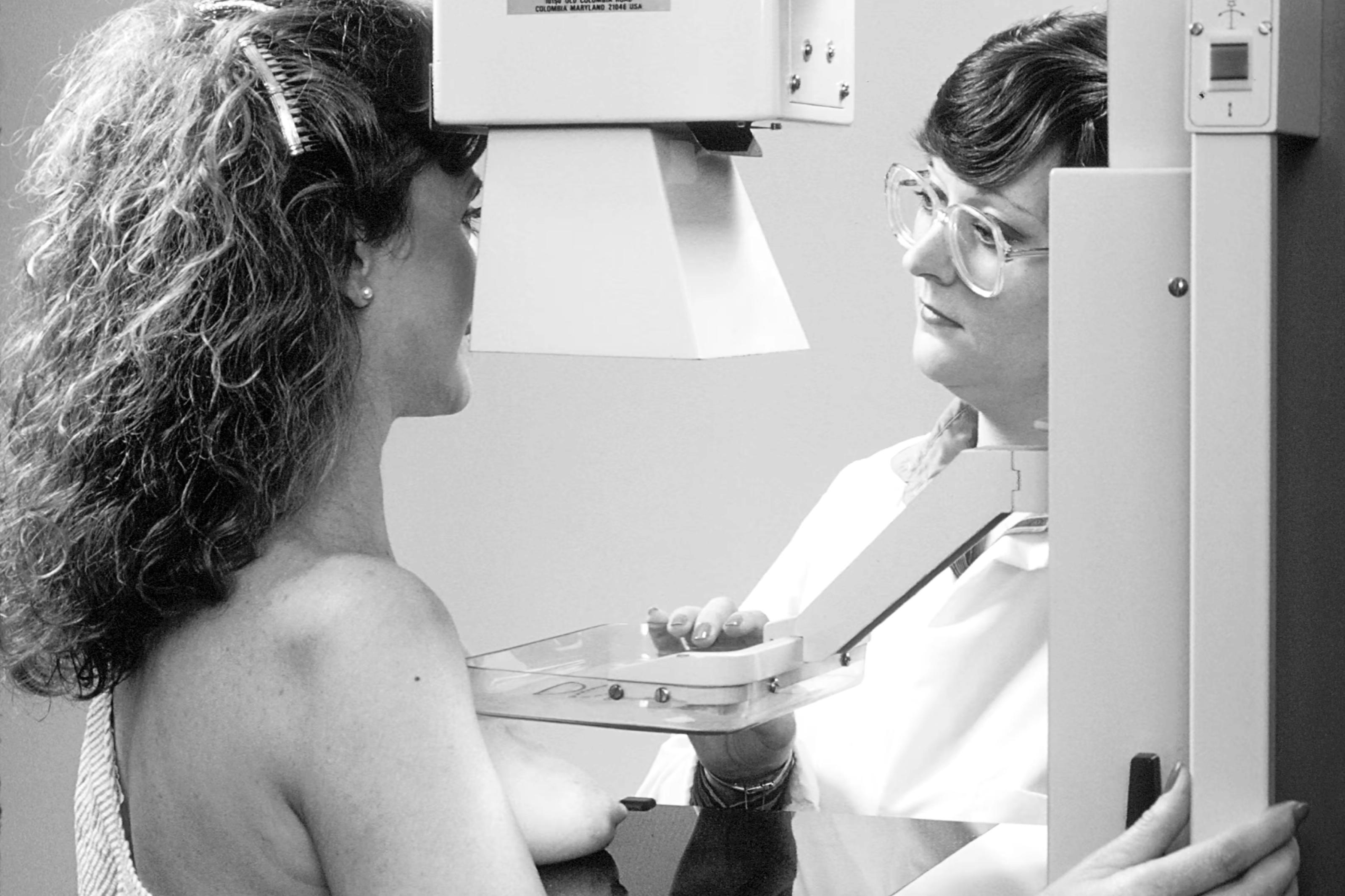
“I’ve heard mammograms cause cancer.” It is one of the most common reasons women give for putting off screening, and it is almost always said with real conviction.
It deserves a real answer rather than an eye roll, because the concern isn’t irrational. Mammograms do use radiation, and radiation is something we are all taught to take seriously. The question is whether the amount involved actually poses the risk people fear.
So let’s take it apart honestly: where the idea came from, what the evidence says, and what the real limitation of mammography actually is (spoiler, it isn’t radiation).
Where the “mammograms cause cancer” idea comes from
The logic is easy to follow, which is exactly why it spreads.
A mammogram is an X-ray, and X-rays are a form of ionizing radiation. High doses of ionizing radiation, the kind involved in industrial accidents or repeated high-dose medical exposure, can damage DNA and raise cancer risk over time. Put those two facts side by side, skip the part about dose, and you land on a conclusion that sounds airtight: a screening test that uses radiation must be causing the thing it looks for.
The missing piece is dose. In radiation, the amount is the entire story, and the amount in a modern mammogram is nothing like the scenarios that make radiation dangerous.
There is also a history here. Mammography was first performed in 1913 (MDPI Healthcare Journal, 2023). The imaging technology of a century ago has almost nothing in common with the equipment used today, but the reputation of “old X-ray machines” has outlived the machines themselves.
So, do mammograms cause cancer? Are mammograms safe?
Here is the direct answer, without hedging.
There is a lot of information, and misinformation, circulating about breast imaging. The radiation exposure from a single mammogram is very low, roughly equivalent to the natural background radiation from a few weeks of daily life. Major medical organizations including the American Cancer Society and the FDA continue to support mammography as an important screening tool.
That last part matters. These are not organizations with a reason to be casual about radiation safety. They are the same bodies that regulate imaging equipment and set screening guidelines, and they have looked at this question far more rigorously than any social media thread. Their position has not wavered: for women at average risk, the benefit of finding breast cancer early outweighs the very small exposure involved.
How much radiation is in a mammogram, really?
If you are searching how much radiation is in a mammogram, the useful comparison isn’t to a nuclear accident. It is your ordinary Tuesday.
You are exposed to background radiation constantly, from the ground beneath you, the air around you, and the sky above you. It is simply part of living on this planet. A single mammogram lands in roughly the same neighborhood as a few weeks of that ordinary, unavoidable exposure.
That is the context that almost never makes it into the conversation. The comparison isn’t “radiation versus no radiation.” It is “a few extra weeks’ worth of what you are already living in, once a year, in exchange for the chance to find something early.”
Myth vs. reality, at a glance
What you may have heard
What is actually true
“Mammograms cause cancer.”
The radiation exposure from a single mammogram is very low, roughly equivalent to the natural background radiation from a few weeks of daily life. Major medical organizations including the American Cancer Society and the FDA continue to support mammography as an important screening tool.
“Ultrasound is the safer replacement.”
Ultrasound is radiation-free, but it is supplemental. For women at average risk it is not considered a stand-alone substitute for a screening mammogram.
“Thermography is a radiation-free alternative.”
Breast thermography is not FDA-approved for breast cancer screening, and it is not considered a replacement for or equivalent to mammography or ultrasound by major medical organizations including the FDA and the American College of Radiology.
“A normal mammogram means I am in the clear.”
Usually reassuring, but not the full picture if you have dense tissue. In extremely dense breasts, mammograms may miss up to 50% of the cancers that are present (Kolb et al., Radiology 2002; FDA, March 2023).
The real limitation of mammograms isn’t radiation. It’s dense tissue.
Here is the part that gets far less attention than it should, and it is the thing actually worth your energy.
On a mammogram, dense fibroglandular tissue shows up white. Tumors also show up white. In a dense breast, looking for a small cancer can be like trying to spot a snowflake in a snowstorm. In extremely dense breasts, mammograms may miss up to 50% of the cancers that are present (Kolb et al., Radiology 2002; FDA, March 2023).
And this is not a rare situation. Nearly half of women over 40 have dense breast tissue (National Cancer Institute, 2024). Women with dense breast tissue are also four to six times more likely to develop breast cancer than women with non-dense breasts (Boyd et al., NEJM 2007).
The FDA takes this gap seriously enough that, since September 10, 2024, every woman must be told her breast density after her mammogram. Not because mammograms are dangerous, but because they have a known blind spot, and you deserve to know whether you are standing in it.
So if you are going to worry about something, worry about the thing that is actually documented: not that your mammogram is harming you, but that in dense tissue it may not be showing you everything.
“Can I get an ultrasound instead of a mammogram?”
This is usually the next question, and the answer is an honest no, at least not as a swap.
For women at average risk, breast ultrasound is not considered a stand-alone substitute for a screening mammogram. The real breast ultrasound vs. mammogram question isn't which one to pick, it's whether you need both. The two tools are good at different things. Mammography is exceptional at picking up microcalcifications, the tiny calcium deposits that can be one of the earliest signs of cancer, and it is the most studied breast imaging tool in existence. Ultrasound is less reliable for those. What ultrasound does well is see through dense tissue, exactly where a mammogram struggles.
That is why the goal isn’t either/or. It is layering. When added to mammography, automated breast ultrasound detects 35.7% more breast cancers than mammography alone (FDA PMA P110006). Not instead of. Added to.
It is also worth saying plainly: no screening tool detects 100% of breast cancers, and ultrasound is no exception. What supplemental screening does is improve the chance that something is found early, which is the whole point. When breast cancer is caught at the localized stage, the five-year survival rate is over 99% (American Cancer Society, Cancer Facts & Figures 2026).
Does a breast ultrasound use radiation?
No. Ultrasound uses sound waves, the same safe technology used to image babies during pregnancy. There is no ionizing radiation, which is why it is considered safe for repeated use (RadiologyInfo.org, RSNA/ACR; U.S. FDA). There is also no compression and no contrast dye.
For women who are anxious about radiation, that fact tends to land hard, and it is a legitimate reason to add a radiation-free layer to your screening. It is not a reason to drop the mammogram that ultrasound is meant to support.
What to actually do with this
If the fear has been keeping you from screening, here is the version that respects both your concern and the evidence:
- Keep your mammogram. If you are 40 or older and at average risk, it remains the foundation. The radiation concern does not hold up, and skipping it trades a very small exposure for a much larger unknown.
- Find out your density. It is on your report now, by law. It tells you how well your own screening can see.
- Know your personal risk. A validated tool like the Tyrer-Cuzick model estimates your individual lifetime and 10-year risk using your age, family history, and other factors. It takes a few minutes and gives everything else context.
- Ask about supplemental screening. If your tissue is dense, an added, radiation-free layer is FDA-approved for exactly that reason.
A radiation-free supplemental scan, in Los Angeles
BeSound is a breast imaging platform that gives you access to FDA-approved automated breast ultrasound (ABUS), the first FDA-approved supplemental ultrasound screening technology specifically designed for detecting cancer in dense breast tissue, and available for women under 40 who want proactive screening as well.
It is radiation-free, compression-free, and dye-free: about 20 minutes, warm gel and light pressure, and an all-female sonographer team at our Los Angeles location on Melrose Avenue. Your images are reviewed by a board-certified physician, with AI-assisted reading flagging areas for closer review along the way, and a physician always makes the final assessment. Results land in your secure portal within 24 to 48 weekday hours. Pricing is upfront: $349, with HSA and FSA accepted.
It is supplemental, working alongside your mammogram rather than instead of it, because that is what the evidence supports and because you deserve straight answers rather than a scare.
If you have been putting off screening because of something you read, you do not have to keep sitting with it. Book your scan in Los Angeles, or start by learning your personal risk for free.
Good information beats a scary headline every time. You deserve both the truth and the clarity that comes with it.
Not medical advice. Consult a medical professional with any medical questions. BeSound provides supplemental breast ultrasound imaging and does not replace mammography or medical care. Imaging is interpreted by licensed physicians. For marketing purposes only. The American Cancer Society recommends that women at average risk begin regular mammograms at age 40. BeSound is a technology platform that connects individuals with independent medical groups that provide breast imaging services; BeSound is not a healthcare provider.
Read more



Start your BeSound journey
Book your scan and view your results directly in your BeSound dashboard.
Learn your risk
Not sure where to start? Take our free risk assessment to better understand your personal risk factors.