Back to Blog
July 7, 2026
Dense Breast Tissue, Explained: What Your Mammogram Report Is Really Telling You
Dense breast tissue is common, normal, and not a diagnosis. Here is what your mammogram report really means and how to build a screening plan that fits your body.
Share on:
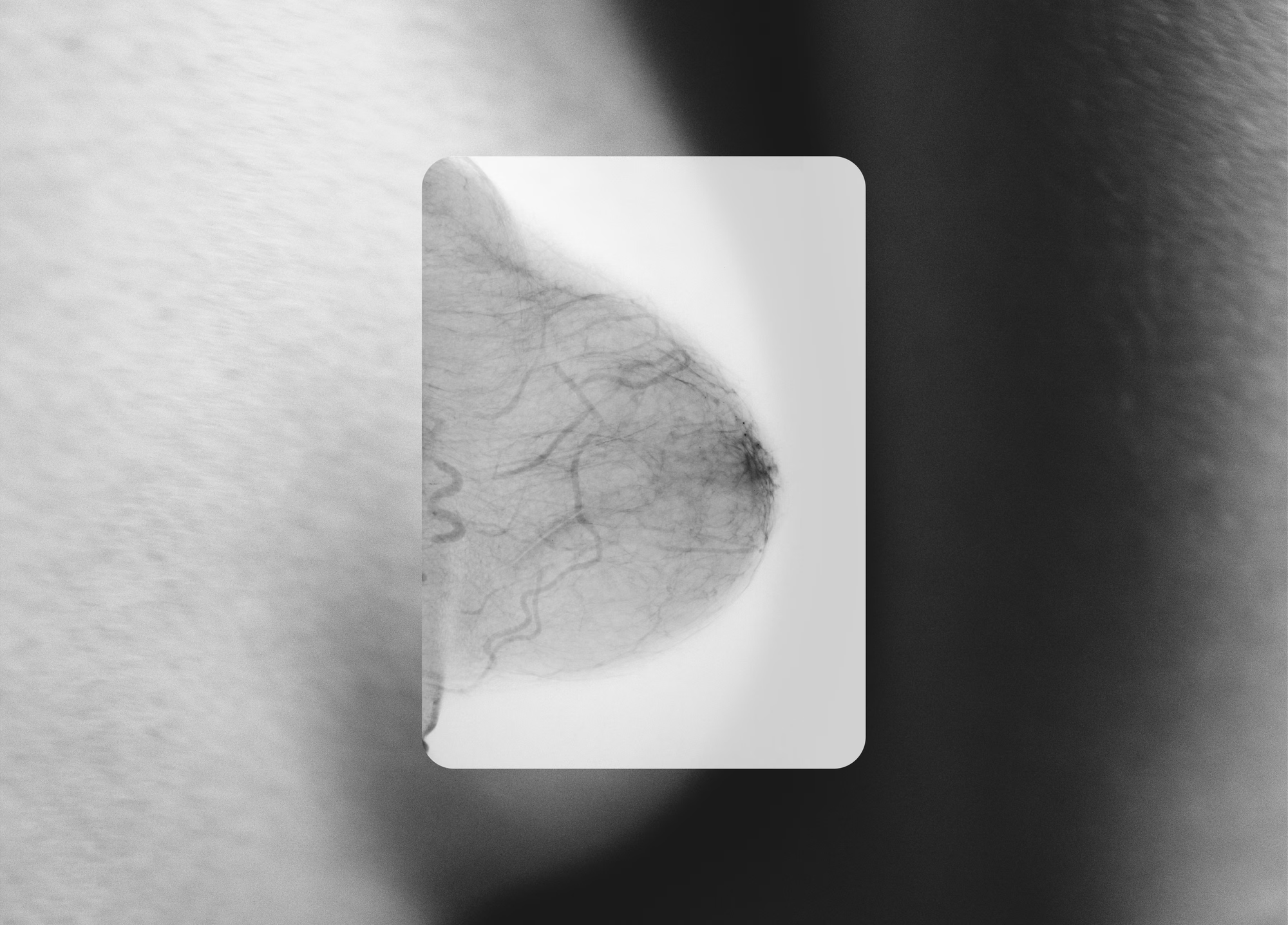
You did the responsible thing. You went in for your mammogram, you held your breath, and a few days later a letter or portal message arrived saying you have dense breast tissue.
Then the questions started. What does that even mean? Did they find something? Should you be worried? And why does it feel like everyone’s breasts are being graded on a scale nobody explained to you?
Take a breath. Dense breast tissue is common, it’s normal, and it is not a diagnosis. But it is something worth understanding, because it changes how well a mammogram can see. Knowing that puts you back in control of your own screening. Let’s walk through it together.
What does dense breast tissue mean?
Your breasts are made of three things: fatty tissue, fibrous (connective) tissue, and glandular tissue, the parts that produce milk. When a radiologist looks at your mammogram, they’re essentially looking at the ratio between fatty tissue and everything else.
The fibrous and glandular tissue together are called fibroglandular tissue, and the more of it you have relative to fat, the “denser” your breasts are said to be. That’s where the term fibroglandular density comes from. It’s simply the clinical way of describing how much of your breast is made up of that dense, active tissue versus soft fat.
Density is not about how your breasts look or feel from the outside. You can’t tell you have dense breasts by touch, and neither can your doctor during an exam. It’s only visible on imaging. Which is exactly why that report language can feel so surprising: it’s describing something you had no way of knowing on your own.
Decoding your report: the density categories
Radiologists sort breast density into four levels (part of a system called BI-RADS). You’ll likely see one of these phrases:
- Almost entirely fatty: very little dense tissue.
- Scattered areas of fibroglandular density: mostly fat, with some dense areas. This is where the phrase scattered fibroglandular density on your report comes from.
- Heterogeneously dense: a lot of dense tissue spread throughout, which can hide small masses. If your report says heterogeneously dense breast tissue, this is the category you’re in.
- Extremely dense: the densest category, where dense tissue makes up most of the breast.
The bottom two categories, heterogeneously dense and extremely dense, are what doctors mean when they say you simply have “dense breasts.” If that’s you, you’re in very good company: nearly half of women over 40 fall into one of these two groups.
What causes dense breast tissue?
There’s no single cause, and to be clear: nothing you did caused it. Breast density is largely determined by factors outside your control:
- Genetics. Density runs in families, the same way body type does.
- Age. Breasts often become less dense over time, particularly after menopause, though many women stay dense their whole lives.
- Hormones. Higher-hormone phases of life (and of your monthly cycle) tend to make tissue denser. Hormone therapy can too.
- Body composition. Women with a lower body weight often have proportionally more dense tissue.
So if you’ve been quietly wondering what causes dense breast tissue and whether it’s a sign you should have done something differently, let that go. It’s a normal variation in human anatomy, like having thick hair or a particular eye color.
Are there symptoms of dense breasts?
This is one of the most-searched questions, so let’s be direct: no. There are no symptoms of dense breasts. Density causes no pain, no lump you can feel, and no outward change. It’s invisible without imaging.
That’s precisely why the topic matters so much. A condition you can’t feel, that affects how well your screening works, is one you can only manage with information. That’s what this article, and honest reporting laws, are here to give you.
Should I worry about dense breast tissue?
Here’s the honest, two-part answer.
No, in the sense that dense tissue itself is not cancer and does not mean something is wrong with you right now.
But yes, it’s worth paying attention to, for two reasons.
First, dense breasts are a recognized risk factor. Women with dense breast tissue are four to six times more likely to develop breast cancer than women with non-dense breasts (Boyd et al., NEJM 2007). Still, it’s a risk factor, not a verdict, and just one of many your doctor weighs.
Second, and more important for your day-to-day peace of mind: dense tissue makes cancer harder to see on a mammogram. On the image, dense fibroglandular tissue shows up white. Tumors also show up white. So a small cancer in dense tissue can be like trying to spot a snowflake in a snowstorm. In extremely dense breasts, mammograms may miss up to 50% of the cancers that are present (Kolb et al., Radiology 2002; FDA, March 2023).
That’s not a knock on mammograms. They remain essential and save lives. It’s simply a known limitation of the technology in dense tissue, and it’s the reason the FDA now requires that women be told their density. You deserve to know how well your own screening can see.
Can dense breast tissue turn into cancer?
No. Dense tissue does not “turn into” cancer, and it isn’t a pre-cancerous condition. The two are separate. What’s true is that having dense breasts is one factor that can raise your overall risk, and it can make an existing cancer harder to detect early. Those are the two real issues, and both are manageable once you know where you stand.
What to do if you have dense breasts
Knowing your density isn’t meant to scare you. It’s meant to help you and your provider build a screening plan that fits your body. A few proactive steps:
- Keep getting your mammograms. If you’re 40 or older, mammography is still the foundation. Nothing here replaces it.
- Know your personal risk. A validated tool like the Tyrer-Cuzick model estimates your individual lifetime and 10-year risk using your age, family history, and other factors. It takes a few minutes and gives your screening plan real context.
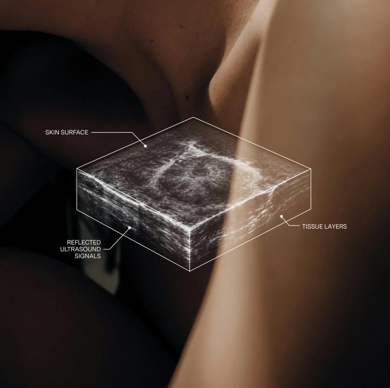
- Ask about supplemental screening. For dense tissue, an added layer of imaging, most often ultrasound, can improve detection where a mammogram falls short. When added to mammography, automated breast ultrasound detects 35.7% more breast cancers than mammography alone (FDA PMA P110006).
That third point is where modern imaging has quietly leapt ahead, and it’s the part most women are never told about.
A clearer picture, for women in Los Angeles
At BeSound, this is the whole reason we exist. Dense breasts aren’t the problem. The century-old technology is. Our Los Angeles locations offer advanced automated breast ultrasound, a comfortable, radiation-free, compression-free scan that is FDA-approved for supplemental screening in women with dense breast tissue, exactly where mammograms struggle. It takes about 20 minutes, and your images are reviewed by a board-certified physician, with results in your secure portal in 24 to 48 weekday hours.
It’s supplemental, working alongside your mammogram rather than instead of it, and it’s built to give you what you came for: answers you can trust.
If you’ve just learned you have dense breast tissue and you’re in the LA area, you don’t have to sit with the uncertainty. Book your scan in Los Angeles, or start by learning your personal risk for free. You deserve imaging that works for your body, and clarity you can hold onto.
Not medical advice. Consult a medical professional with any medical questions. BeSound provides supplemental breast ultrasound imaging and does not replace mammography or medical care. Imaging is interpreted by licensed physicians. For marketing purposes only. BeSound is a technology platform that connects individuals with independent partner medical groups that provide breast imaging services; BeSound is not a healthcare provider.
Read more
Start your BeSound journey
Book your scan, understand your lifetime risk, and view your results directly in your BeSound dashboard.
Join the waitlist
Our modern breast imaging experience is coming to more cities across the U.S. Join the waitlist to be the first to know when we launch in your city.
Learn your risk
Our free assessment gives results based on your unique health profile using a clinically validated model trusted by healthcare providers.